
Contents
What varicose veins are and how they form
Signs, symptoms, and who is at higher risk
Complications that require attention
Diagnosis: clinical exam and venous Doppler ultrasound
CEAP classification in simple language
Treatment: from conservative care to procedures
Pregnancy and varicose veins
Prevention in everyday life
Myths and facts
Quick FAQ
Health disclaimer
References and recommended reading
What varicose veins are and how they form
Varicose veins are dilated, tortuous superficial veins, most commonly in the legs. The core problem is valve failure. When venous valves do not close properly, blood refluxes downward, pressure rises, and the vein dilates. This is chronic venous insufficiency. Beyond visible varicose veins, chronic venous disease also includes swelling, skin changes, and—at advanced stages—ulcers.
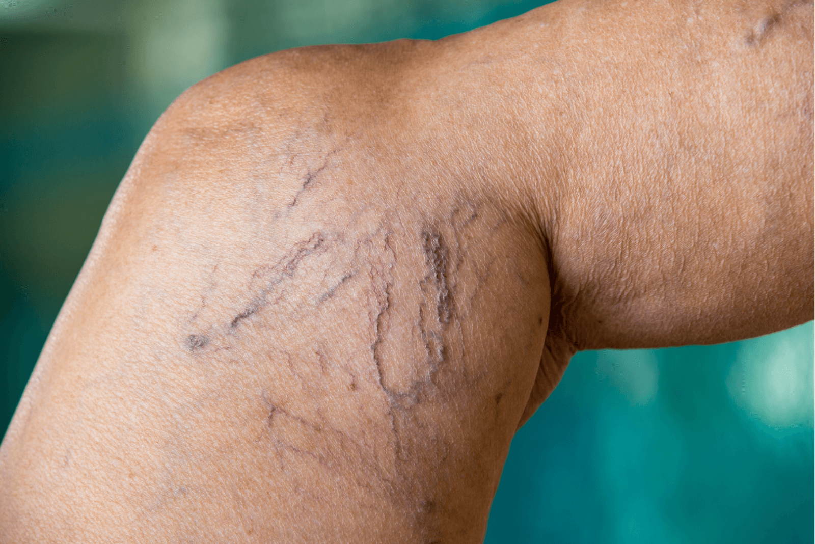
Not every visible vein is a varix. Telangiectasias and reticular veins (“spider veins”) are smaller and more superficial. They may be symptomatic or cosmetic, but share the same physiology of venous overload.
Signs, symptoms, and who is at higher risk
Typical symptoms
Heaviness, aching, or burning pain in the legs, worse at day’s end
Ankle and leg edema that improves with leg elevation
Night cramps, tingling, itching
Skin darkening and hardening around the ankle in advanced stages
Risk factors
Strong family history
Age and female sex
Pregnancy and hormonal exposure
Obesity and sedentary lifestyle
Jobs with long periods standing or sitting without breaks
Prior thrombosis or venous trauma
Complications that require attention
Superficial thrombophlebitis: painful, reddened cord along a vein
Bleeding from superficial varices after minor trauma
Venous eczema, lipodermatosclerosis, and venous ulcers near the medial malleolus
Deep vein thrombosis (DVT): sudden unilateral swelling and pain, especially after immobilization; this is an emergency
Diagnosis: clinical exam and venous Doppler ultrasound
Assessment starts at the visit, ideally with the patient standing. The test that confirms reflux and maps venous anatomy is venous Doppler ultrasound (duplex scan). It identifies which veins are incompetent, the direction of flow, and any thrombi. This map guides a personalized plan—different patterns need different approaches.
CEAP classification in simple language
CEAP describes clinical severity and etiology:
C0–C2: no visible signs, spider veins, or true varicose veins
C3: edema
C4: skin changes such as hyperpigmentation and induration
C5: healed ulcer
C6: active ulcer
“E” covers extent, “A” the anatomy, and “P” the pathophysiology. Practically, higher C means greater urgency to treat and prevent progression.
Treatment: from conservative care to procedures
Goals: reduce symptoms, prevent complications, and improve function and appearance. Plans often combine lifestyle measures and, when indicated, targeted procedures.
Conservative measures
Graduated compression stockings (often 15–20 or 20–30 mmHg), put on in the morning
Daily leg elevation for a few minutes
Calf-muscle exercise, walking, cycling
Weight management and avoiding long periods without movement
Skin hydration and treatment of associated dermatitis
Venoactive agents (e.g., micronized flavonoids, horse chestnut) may ease symptoms, but do not correct reflux
Minimally invasive procedures
Choice depends on ultrasound mapping, vein diameter, and whether the saphenous vein or tributaries are incompetent.
Liquid sclerotherapy: sclerosant injections for spider and small reticular veins
Foam sclerotherapy (e.g., polidocanol foam): useful for larger segments and tortuous varices
Endovenous thermal ablation
Endovenous laser
Radiofrequency ablation
These seal an incompetent saphenous vein from within. Typically done with local anesthesia and quick return to activities.
Non-thermal techniques: cyanoacrylate closure or mechanochemical ablation for selected profiles
Microphlebectomy (ambulatory phlebectomy): removal of varicose segments through tiny incisions, often combined with other techniques
Conventional surgery (stripping): remains an option when minimally invasive methods are unavailable or anatomy is unsuitable
After a procedure
Short course of compression, early walking, and rapid return to routine. Bruising, pigmentation, and a “cord-like” feeling can occur and usually resolve. Follow-up ultrasound confirms success and guides touch-ups if needed.
Pregnancy and varicose veins
Pregnancy increases blood volume and relaxes venous walls. Varicose veins often worsen in later trimesters and improve postpartum. Focus on compression stockings, light activity, and leg elevation. Procedures are usually deferred until after pregnancy unless there is a complication. For long trips, hydrate, walk every 1–2 hours, and use compression to reduce DVT risk.
Prevention in everyday life
Activate calf muscles whenever standing still
Walk during work breaks and stretch regularly
Avoid very tight garments that compress the groin or waist
Manage weight and favor a fiber-rich diet
On long flights or periods of immobilization, use compression and hydrate
Myths and facts
“Crossing your legs causes varicose veins.” Myth. Static posture worsens symptoms but is not an isolated cause.
“Only women get varicose veins.” Myth. Men are affected too and often underdiagnosed.
“Compression stockings are addictive.” Myth. They aid venous return and relieve symptoms.
“Procedures are only cosmetic.” Myth. Treating reflux reduces pain, swelling, and ulcer risk.
Quick FAQ
When should I see a vascular specialist?
If you have pain, swelling, persistent itching, skin darkening, wounds, a painful cord, or bleeding from a superficial vein.
Is ultrasound always necessary?
In overt varicose disease, duplex mapping guides the plan and avoids treating only surface veins when the saphenous trunk is the source.
Do varicose veins come back?
New segments can appear over time. Ongoing follow-up allows control and touch-ups when needed.
Do compression stockings cure varicose veins?
They relieve symptoms and slow progression, but do not fix damaged valves.
Can I train legs at the gym?
Yes. Strengthening the calves helps. Avoid standing still after intense sets; walk a few minutes.
Health disclaimer
This content is educational and does not replace medical care. Sudden leg pain and swelling, marked asymmetry, shortness of breath, or chest pain require immediate evaluation for possible thrombosis or embolism.
References and recommended reading
NICE. Varicose veins in the legs: diagnosis and management.
ESVS. Clinical practice guidelines on the management of chronic venous disease.
American Venous Forum / Society for Vascular Surgery. Practice guidelines for superficial venous disease and venous ulcer management.
Cochrane Reviews. Compression therapy, foam sclerotherapy, and endovenous ablation.
American College of Phlebology / American Vein & Lymphatic Society. Consensus statements on endovenous therapies and sclerotherapy.